


Learning about the Cranial Cruciate Ligament, and an Introduction to the MRIT Procedure (Modified Retinacular Imbrication Technique or Lateral Fabellar suture)
Understanding the Cranial Cruciate Ligament (CCL) in Dogs
The cranial cruciate ligament (CCL) plays a critical role in the knee—or more properly, the stifle—of a dog. If you’re familiar with sports injuries, you may have heard about the anterior cruciate ligament (ACL) in people. Essentially, the CCL in dogs serves the same purpose as the ACL does in humans: it stabilizes the knee by preventing excessive forward motion and rotation of the lower leg relative to the thigh.
Dogs have two cruciate ligaments within the stifle joint: the cranial and caudal cruciate ligaments. They get their name from the way they cross each other inside the knee, forming an “X,” which is the origin of the term “cruciate.” When the CCL is injured or torn—a common occurrence in active or aging dogs—it results in instability, pain, and lameness, much like an ACL injury would in a weekend soccer player or skier.
Signs and Consequences of a Torn CCL
A torn cranial cruciate ligament (CCL) in dogs is more than just a minor setback—it’s a game-changer for your canine companion’s mobility. Typically, a dog with a CCL injury will show obvious lameness, often struggling to walk or rise from a resting position. You might notice an unsteady, limping gait, and sometimes, an awkward shuffle that just looks “off.”
Beyond the limp, the main issue is instability in the knee joint itself. When the CCL is damaged, the shin bone (tibia) is able to slide too far forward relative to the thigh bone (femur). This unchecked movement not only worsens the limp but also accelerates wear and tear on the joint.
If left untreated, the instability leads to a cascade of consequences:
- Progressive joint pain
- Ongoing inflammation
- Damage to joint cartilage
- Development of osteoarthritis (OA)
Over time, even a stoic dog may become reluctant to bear weight on the affected leg and may show signs of muscle loss in the thigh. In other words, a torn CCL doesn’t just slow your dog down for a few days—it can set the stage for chronic pain and permanent mobility problems if not addressed.
What Happens When the Cranial Cruciate Ligament Tears?
When the cranial cruciate ligament (CCL) in a dog’s knee is damaged, the stifle joint loses its normal stability. As a result, the tibia (shin bone) shifts abnormally forward in relation to the femur (thigh bone). This excessive movement—often detected as a “drawer sign” during examination—compromises the dog’s ability to walk comfortably and leads to pain and lameness.
Over time, this instability not only affects mobility but also places extra strain on the cartilage and joint surfaces, accelerating the development of arthritis. If left untreated, even routine activities like walking or climbing stairs become increasingly difficult, and degenerative joint changes progress.
Why Is Surgery Often Needed for a Torn Cruciate Ligament?
When a dog’s cranial cruciate ligament (CCL) is torn, the knee joint becomes unstable, making it difficult and often painful to walk or run—think of it as a chair with a missing leg: it just can’t support you properly. Particularly in larger or energetic breeds who love their daily jogs and grassy zoomies, this instability can make everyday activities impossible.
Surgical stabilization is usually the best way to restore proper joint function for these dogs. Acting quickly helps prevent further damage inside the knee, such as cartilage wear and worsening arthritis. Ultimately, early surgical intervention doesn’t just reduce discomfort; it gives your dog their best shot at a smooth recovery and a return to their usual tail-wagging adventures.
Choosing the Right Surgical Option
When it comes to treating a torn CCL, your veterinarian is a key partner in selecting the most appropriate surgical approach for your dog. After a thorough orthopedic examination and review of X-rays or other diagnostics, your veterinarian will consider several factors:
- The size and weight of your dog
- Your dog’s age and activity level (weekend hiker vs. Couch potato)
- The presence of other stifle (knee) issues, such as arthritis or meniscal injury
- Any concurrent health problems that might affect recovery
Based on this information, your vet will discuss the pros and cons of available surgical techniques—such as MRIT (as described above). They’ll help you weigh factors like expected recovery time, long-term outcomes, and potential complications. This collaborative process ensures the chosen repair aligns with both your dog’s needs and your lifestyle.
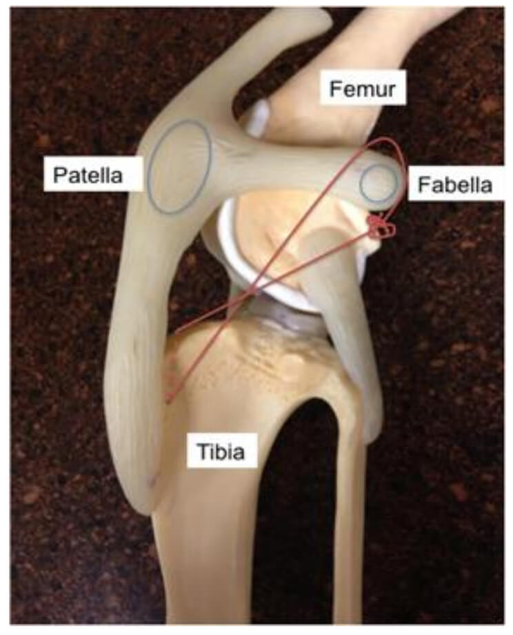
MRIT Procedure
The MRIT procedure involves an incision over the inside or outside aspect of the knee (surgeon dependent). The joint is explored (arthrotomy) to examine the CCL, assess arthritis, and look for any meniscal injury. If there is a meniscal injury, the meniscus is partially removed. An intact meniscus may also be surgically “released” as a prophylactic measure against future injury (surgeon dependent). Next, a sterile suture is selected depending on the size, age, and activity level of the patient. The suture is passed around the fabella and through a tunnel created in the tibia. The suture is then tightened to stabilize the knee. See figure below. For larger patients, a suture may be placed on both the medial and lateral aspect of the knee.
Post-Operative Care
Post-operative care after any extra-capsular stabilization includes activity restriction, incision care, physical therapy, and specific medications.
Activity Restriction – The post-operative recovery period usually lasts for about 8 weeks. During this period, the patient needs to have their activity restricted as to not cause complications with the stabilization. Too much activity can lead to implant failure, meniscal injury, and pain. We usually recommend confinement (crate, kennel, enclosure, small room), leash walks only, no jumping, no playing, no climbing, and no running for the majority of the recovery period. As the patient recovers, the surgeon will implement gradual return to normal activity.
Incision Care – It usually takes about 2-3 weeks for the incision and soft tissues to heal. During this time, it is important to monitor the incision for any excessive swelling, oozing, or incisional dehiscence (opening up). We also recommend an E-collar (cone) placed around the dogs’ head in order to keep them from chewing or licking at the incision. Licking or chewing at the incision can lead to dehiscence and/or infection of the site, which can be a serious complication especially if infection reaches the implant.
In addition, cold and/or warm compress may be implemented to decrease incisional swelling.
Pain Management
Pain management during and after stifle surgery is critical. Be sure to give all medications as prescribed and continue them until they are gone, unless directed otherwise by your veterinarian. Pain relief helps keep your pet comfortable and is essential for optimal healing.
Physical Rehabilitation
Physical rehabilitation post-operatively can improve healing and speed up the time it takes your dog to return to their normal activities. Simple, gentle exercises and controlled leash walks, as recommended by your veterinary team, can help restore strength and mobility. Ask your veterinarian about incorporating rehabilitation into your dog’s recovery plan, as tailored exercises may greatly enhance recovery.
By carefully following our post-operative instructions provided, and keeping up with follow-up appointments, you can help ensure the best possible recovery for your pet after the MRIT procedure.
Risks and Success Rates of Extracapsular Repairs
Both traditional extracapsular stabilization techniques and newer procedures are considered external repairs for cranial cruciate ligament (CCL) injuries. The two primary risks associated with these surgeries are infection and failure of the repair. While these risks exist, the success rates are encouraging: at least 85–90% of smaller dogs experience positive outcomes, and the overall complication rate remains relatively low, typically reported between 5% and 8%.
Paying close attention to activity restriction and incision care during recovery is key to minimizing these risks and promoting the best possible surgical outcome.